MR Coils / Visual Cortex Phased-Array MRI Coil
Visual Cortex Phased-Array MRI Coil
Questions about the Güdform 32?
![]()
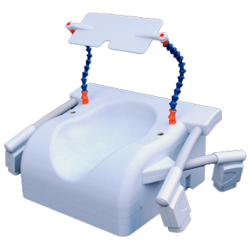
The Güdform 32 channel MR coil is atlas-optimised for visual cortex imaging and provides 7T-like performance on standard 3T scanners.
In many locations, the SNR of this posterior-only coil is more than 2X greater than a typical vendor-provided whole-head 32 channel coil, and allows for fast, high-resolution imaging of the visual cortex, down to 0.75mm isotropic voxel resolution for fMRI, and 0.35mm isotropic resolution for anatomical imaging.
Guide Price: £75000
SKU: P0136


Selected References
Dense, shape-optimized posterior 32-channel coil for submillimeter functional imaging of visual cortex at 3T.
Farivar R., Grigorov, F., van der Kouwe A.J., Wald, L.L., and Keil, B. (2015). Magn Reson Med. 2016 Jul;76(1):321-8. doi: 10.1002/mrm.25815
Abstract
PURPOSE:
Functional neuroimaging of small cortical patches such as columns is essential for testing computational models of vision, but imaging from cortical columns at conventional 3T fields is exceedingly difficult. By targeting the visual cortex exclusively, we tested whether combined optimization of shape, coil placement, and electronics would yield the necessary gains in signal-to-noise ratio (SNR) for submillimeter visual cortex functional MRI (fMRI).
METHOD:
We optimized the shape of the housing to a population-averaged atlas. The shape was comfortable without cushions and resulted in the maximally proximal placement of the coil elements. By using small wire loops with the least number of solder joints, we were able to maximize the Q factor of the individual elements. Finally, by planning the placement of the coils using the brain atlas, we were able to target the arrangement of the coil elements to the extent of the visual cortex.
RESULTS:
The combined optimizations led to as much as two-fold SNR gain compared with a whole-head 32-channel coil. This gain was reflected in temporal SNR as well and enabled fMRI mapping at 0.75 mm resolutions using a conventional GRAPPA-accelerated gradient echo echo planar imaging.
CONCLUSION:
Integrated optimization of shape, electronics, and element placement can lead to large gains in SNR and empower submillimeter fMRI at 3T. Magn Reson Med 76:321-328, 2016. © 2015 Wiley Periodicals, Inc.